 Medicine Innovates Medicine Innovates: Delivering innovations in medicine to the world for better health and prosperity

Significance
Atypical megakaryocytes accumulate in myeloproliferative neoplasms and release profibrotic signals that drive collagen deposition, disturb normal hematopoiesis, and contribute to progressive marrow fibrosis. In BCR::ABL-negative myeloproliferative neoplasms, including polycythemia vera, ET, and primary myelofibrosis, mutations in JAK2, MPL, or CALR produce cytokine-independent JAK/STAT signaling. Primary myelofibrosis carries a particularly severe disease burden, with extramedullary hematopoiesis, inflammatory cytokine production, cytopenias associated with marrow fibrosis, splenomegaly, and progression to acute myeloid leukemia. Megakaryocytes are closely connected to that biology: abnormal forms accumulate in diseased marrow, secrete factors such as TGF-β that promote collagen deposition, and can directly shape the fibrotic state.
JAK inhibitors such as ruxolitinib remain central to treatment because they reduce symptom burden and spleen volume. Disease biology, however, extends beyond JAK/STAT signaling alone. Fibrosis and mutant allele burden can persist, and responses to ruxolitinib commonly diminish over time. Experimental depletion of megakaryocytes in MPN models has shown that reducing this lineage can alleviate major features of myelofibrosis, giving a biological basis for approaches directed more specifically at abnormal megakaryocyte development. Previous efforts had also explored altering the maturation of abnormal megakaryocytes, reinforcing the idea that this lineage is not simply a histologic feature of disease but an active participant in its progression. The unresolved question was which molecular dependency could be used to act on megakaryocyte progenitors at a stage capable of influencing the broader disease process.
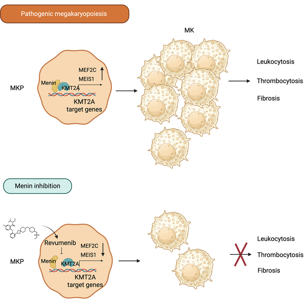
Menin provided an unexpected entry point. The protein associates with KMT2A in transcriptional control of genes such as MEIS1 and HOXA9 and is already a pharmacologic target in molecularly defined acute leukemias. Clinical experience with the menin inhibitor revumenib had also produced decreased platelet counts in a subset of heavily pretreated patients. Rather than treating that observation simply as a hematologic drug effect, the investigators used it to ask a mechanistic question with direct relevance to MPN biology: could menin activity be required for megakaryocyte development, especially for the highly proliferative megakaryocyte progenitors that expand in myelofibrosis?
Jeremy Wen, Anitria Cotton, Rashid Mehmood, Man Mohan, Trent Hall, Amber Broadhurst, Kevin Zhang, Yunusa Olufadi, Guolian Kang, Bridget Marcellino, John Mascarenhas, Ronald Hoffman, Laura Janke, Sandeep Gurbuxani, Gerard McGeehan, and John Crispino from St. Jude Children’s Research Hospital developed a pharmacologic and genetic strategy for targeting menin-dependent megakaryocyte progenitors in myeloproliferative neoplasms. The approach combines menin inhibition with MEN1, MEF2C, and MEIS1 perturbation to define the transcriptional dependency of abnormal megakaryopoiesis. They established revumenib as an intervention acting on proliferative megakaryocyte progenitors across human cultures, patient-derived cells, and several MPN mouse models. They also defined a mechanistically complementary pairing with ruxolitinib that addresses menin-dependent progenitor programs alongside JAK/STAT signaling.
The researchers began with healthy human CD34+ progenitor cells and asked whether pharmacologic menin inhibition altered megakaryocyte production selectively. Revumenib reduced megakaryocyte colony formation, decreased CD41+ megakaryocyte progenitors, suppressed immature and mature megakaryocyte production, reduced polyploidization, and increased apoptotic markers. Myeloid and erythroid colony formation remained largely unaffected in the colony assays. Ziftomenib produced a similar response, and CRISPR disruption of MEN1 reproduced the decrease in megakaryocyte progenitors, megakaryocyte output, ploidy, and cell survival. Using both drug inhibition and gene disruption was scientifically informative because convergence of the two approaches connected the cellular phenotype to menin itself rather than to an isolated compound-specific response.
Single-cell RNA sequencing then resolved where that dependency appeared across differentiating hematopoietic populations. Revumenib and MEN1 loss markedly reduced megakaryocyte progenitor and megakaryocyte populations, accompanied by increased proportions of common myeloid progenitors and megakaryocyte-erythroid progenitors. Within megakaryocyte progenitors, menin inhibition reduced expression of MEIS1, MEF2C, and PBX3. Pathway analysis also identified depletion of programs associated with cell cycle, RNA splicing, and proliferation in the megakaryocytic populations. JAK/STAT pathway programs were not significantly enriched among the affected pathways, separating the transcriptional action of menin inhibition from direct suppression of the canonical signaling axis that drives MPNs. The cellular redistribution seen after either pharmacologic or genetic loss of menin therefore reflected a change concentrated around megakaryocyte progenitor specification or maintenance.
The team next tested revumenib across genetically distinct mouse models. In Jak2V617F disease, treatment reduced abnormal blood counts, spleen enlargement, and marrow reticulin fibrosis, with treated marrow and spleen showing normalized morphology. In an inducible Jak2V617F model, revumenib and ruxolitinib affected overlapping but different disease features, and their combination normalized several blood-count and organ-weight measures. The design was particularly informative because it separated an intervention directed at menin-dependent progenitor biology from established JAK inhibition within the same disease setting.
The MPLW515L myelofibrosis model provided a deeper view of the megakaryocyte-fibrosis relationship. Revumenib reduced thrombocytosis and leukocytosis, decreased abnormal megakaryocyte burden, lowered circulating inflammatory and profibrotic mediators including TGF-β and TNF-α, reduced marrow fibrosis, and extended survival. The MplS504N model produced the same biological direction, including fewer marrow megakaryocytes, more normal megakaryocyte morphology, reduced fibrosis, and restoration of spleen architecture. Primary human material then connected these observations directly to myelofibrosis: revumenib reduced megakaryocyte colony formation, megakaryocyte production, ploidy, and progenitor abundance in samples carrying JAK2, CALR, or combined mutations. In a patient-derived xenograft, treatment reduced human cells in peripheral blood and marrow and decreased human CD34+CD38+ progenitors.
The authors afterward focused on MEF2C and MEIS1. CRISPR loss of MEF2C reproduced the effect of menin inhibition on megakaryocyte progenitors, and restoring MEF2C expression protected progenitor numbers during revumenib exposure. MEIS1 loss also reproduced major features of MEN1 disruption, though restoration experiments distinguished MEF2C as a particularly strong mediator of the progenitor response. These experiments place altered transcriptional control of proliferative megakaryocyte progenitors near the center of revumenib activity rather than treating reduced platelet production as an isolated pharmacologic observation.
The findings of St. Jude Children’s Research Hospital researchers led by Professor John Crispino reaches beyond a reduction in platelet-producing cells. Myelofibrosis is driven in part by an abnormal megakaryocyte compartment that expands and reshapes the marrow environment through profibrotic and inflammatory signaling. By tracing revumenib activity back to proliferative megakaryocyte progenitors, the researchers connect a druggable transcriptional dependency to one of the cell populations that actively sustains the disease phenotype. The reduction in marrow fibrosis across several MPN models fits that mechanism: fewer atypical megakaryocytes occurred alongside lower levels of mediators such as TGF-β and TNF-α and restoration of marrow morphology. The experimental sequence gives the fibrosis response a cellular and molecular explanation rather than leaving it as an isolated histopathologic observation.
A second contribution is the mechanistic separation from JAK/STAT inhibition. Revumenib altered menin-KMT2A target-gene expression without suppressing JAK/STAT pathway activity, and ruxolitinib acts through a different disease axis. Their combined activity in mouse models is consistent with complementary targeting of signaling and megakaryocyte progenitor biology. That distinction matters scientifically because it provides a rationale for combining interventions that act through separate molecular programs rather than duplicating the same target. In the inducible Jak2V617F system, for example, the combination normalized several hematologic and organ measurements, linking molecular complementarity to effects measurable at the whole-animal level.
MEF2C and MEIS1 sharpen the molecular interpretation further. Loss of either gene reproduced substantial parts of the megakaryocyte phenotype caused by MEN1 disruption, but restoration of MEF2C preserved megakaryocyte progenitor numbers during revumenib treatment. The data support MEF2C as a major downstream mediator of menin activity in this lineage and place menin-dependent transcription upstream of progenitor proliferation and differentiation. The response is also not confined to one initiating MPN mutation, since experimental systems incorporating JAK2, MPL, and CALR alterations showed activity in the same general biological direction. This gives the proposed mechanism a degree of coherence across genetically different forms of MPN biology.
For therapeutic development, healthy human progenitors defined lineage selectivity, genetic perturbation established target dependence, single-cell profiling localized the transcriptional response, multiple murine disease models connected that response to fibrosis and blood-count abnormalities, and primary PMF samples showed activity against patient-derived malignant progenitors. The patient-derived xenograft added another level by demonstrating suppression of human hematopoietic cells and progenitors within an in vivo setting. On this experimental basis, the authors propose clinical evaluation of menin inhibitors such as revumenib in myelofibrosis. Their observations also provide a mechanistic basis for studying revumenib together with ruxolitinib, since the agents act on distinct components of MPN biology rather than a single shared molecular process.

Reference
Wen J, Cotton A, Mehmood R, Mohan M, Hall T, Broadhurst AL, Zhang K, Olufadi Y, Kang G, Marcellino B, Mascarenhas J, Hoffman R, Janke LJ, Gurbuxani S, McGeehan GM, Crispino JD. Menin-dependent megakaryocyte proliferation and fibrosis in myeloproliferative neoplasms. Cancer Cell. 2026 ;44(8):1639-1652.e6. doi: 10.1016/j.ccell.2026.06.008.
Go to Cancer Cell