 Medicine Innovates Medicine Innovates: Delivering innovations in medicine to the world for better health and prosperity
Significance
Fluorescence imaging in the second near-infrared window allows biological structures to be seen deeper inside the body, where visible light is strongly absorbed and scattered by tissue. This window covers wavelengths from about 900 to 1700 nm, although some parts of this range provide clearer images than others. Imaging above 1400 nm can show blood vessels more clearly because water in the tissue absorbs much of the scattered light. This improves image contrast, but the fluorophore must still emit enough light at these longer wavelengths to produce a clear signal. The shortage of suitable organic dyes therefore remains a molecular problem as much as an optical one. A fluorophore intended for long-wavelength NIR-II imaging must absorb strongly, emit far enough into the deep-NIR region, and resist photobleaching and chemical degradation under biologically relevant conditions. These properties are difficult to optimize simultaneously. Extending conjugation can shift absorption and emission toward longer wavelengths, but red-shifting a molecular scaffold may reduce fluorescence efficiency. Small Stokes shifts create an additional complication because overlap between absorption and emission promotes self-absorption, diminishing the signal available for imaging. Xanthene fluorophores provide an adaptable structural basis that maybe capable to address these coupled requirements because their optical response can be modified through changes in conjugation, donor strength, substitution pattern, and molecular geometry. However, for optimal design the useful probe must retain adequate brightness and in the same time has sufficiently large separation between excitation and emission, and the molecular structure must also remain stable in the presence of light, changes in pH, biological nucleophiles, and reactive chemical species.
In a recently published research paper in Journal of Materials Chemistry B Dr. Tong Xiangli, Dr. Yani Shang, Dr. Yongjie Chen, Dr. Shenwei Chen, Professor Hongbing Ji, and Professor Jin Li from Zhejiang University of Technology working together with Dr. Qinghua Wu from Anhui University of Chinese Medicine developed a molecular engineering platform that combines benzannulation-driven π-extension with terminal donor modification in an asymmetric xanthene scaffold. The platform produced three tunable deep-NIR fluorophores with distinct absorption, emission, brightness, and Stokes-shift profiles. They also converted the best-performing compound, NIR-842, into DSPE-PEG2000 nanoparticles that retained strong fluorescence and colloidal stability in water.
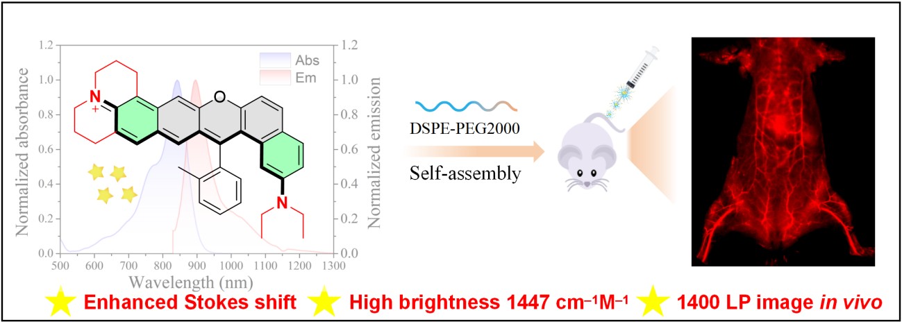
The researchers prepared three asymmetric xanthene fluorophores, NIR-820, NIR-842, and NIR-864 by using a short sequence of chemical reactions. They conducted structural analysis and confirmed that the asymmetric molecule they wanted had formed. The terminal julolidine group was distorted, and the phenyl substituent was rotated relative to the molecular core. This non-planar shape created steric hindrance, limited close molecular stacking, and helped reduce aggregation.
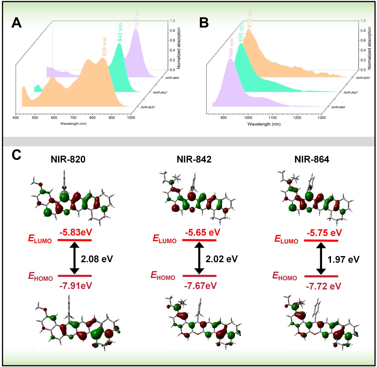
The team examined three dyes were how terminal electronic changes affected their optical behaviour and observed NIR-820 showed deep near-infrared absorption and emission with a large Stokes shift. Replacing its methoxy group with a stronger diethylamino donor produced NIR-842 and shifted both absorption and emission to longer wavelengths. This change strengthened the push–pull electronic character of the molecule, increased intramolecular charge transfer, and greatly improved brightness.
The authors found a further substitution produced NIR-864 and shifted the spectrum still farther toward longer wavelengths. However, this additional red shift was accompanied by a smaller Stokes shift and lower brightness. NIR-842 offered the best balance among emission wavelength, Stokes shift, and fluorescence intensity and was much brighter than the other two dyes.
They also conducted electronic-structure calculations and found the energy gap gradually decreased across the series, which agrees with the progressive red shift in absorption and emission. NIR-842 and NIR-864 also showed stronger charge-transfer character, with the excited-state electron density spread more broadly across the conjugated xanthene structure. The main absorption process was assigned to a transition between the highest occupied and lowest unoccupied molecular orbitals. All three dyes resisted photobleaching more effectively than indocyanine green. They remained stable across the physiologically relevant pH range and showed little reaction with cysteine, glutathione, hydrogen peroxide, or sodium hypochlorite under the tested conditions. Steric protection around the central methine carbon helped limit nucleophilic attack.
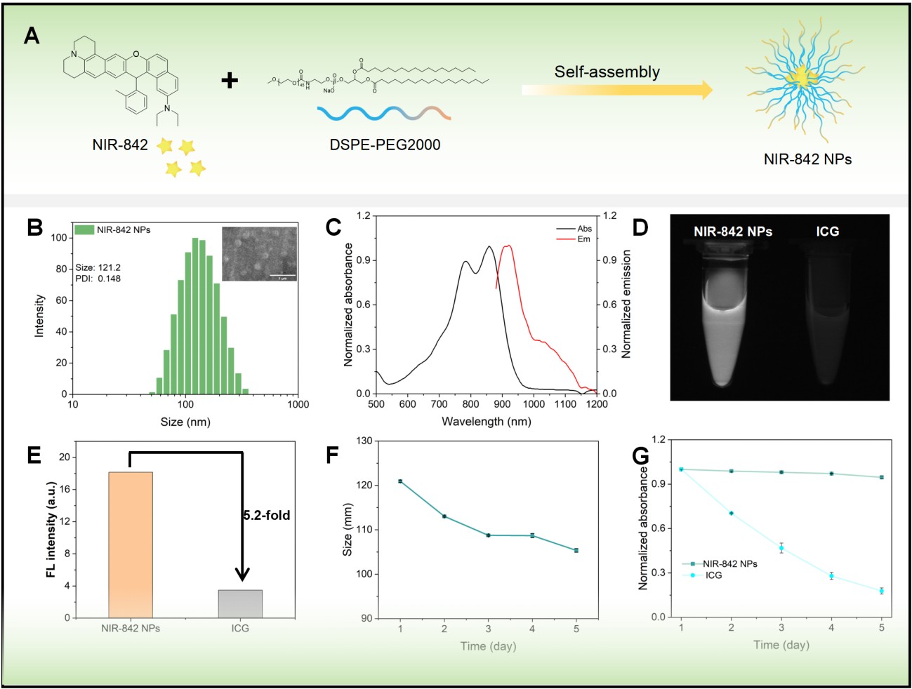
The authors then encapsulated NIR-842 with DSPE-PEG2000 to form uniform spherical nanoparticles and found encapsulation improved its compatibility with water and shifted the optical response slightly toward longer wavelengths. In phosphate-buffered saline, the nanoparticles were much brighter than indocyanine green and remained physically and optically stable during storage in water.
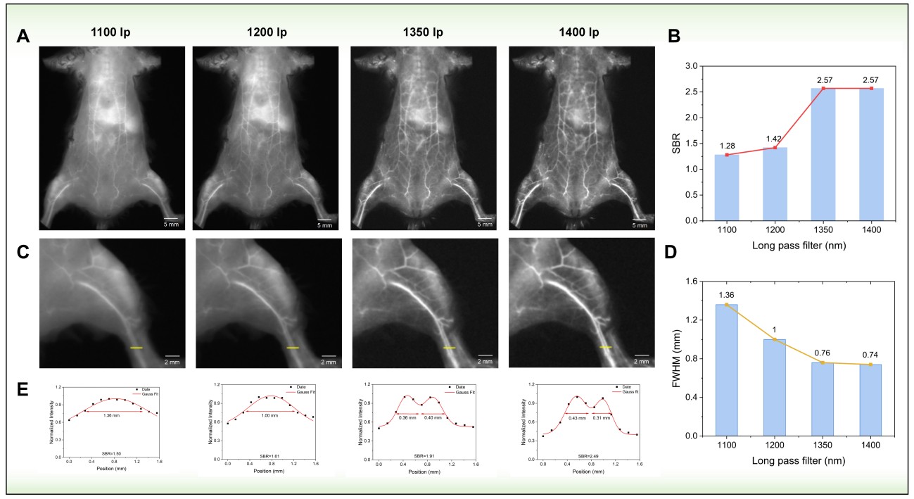
The fluorophore platform developed by Professor Jin Li and colleagues is suited to biomedical imaging situations in which small vascular structures must be visualized through tissue with high contrast and the strongest candidate, NIR-842, combines high brightness, a large Stokes shift, strong photostability, and an emission tail extending beyond 1400 nm. After formulation as DSPE-PEG2000 nanoparticles, it enabled clear imaging of mouse blood vessels under 880 nm excitation, with the best image quality obtained when fluorescence above 1400 nm was collected. This makes the system relevant to real-time angiography, where accurate delineation of vessel boundaries and fine branches is essential.
One immediate application is the non-invasive assessment of superficial and deeper vascular networks. The improved signal-to-background ratio and narrower apparent vessel profiles at longer detection wavelengths could support the visualization of small vessels that are difficult to distinguish using shorter-wavelength NIR-II detection. Such capability may be useful in preclinical studies of vascular development, tissue perfusion, vessel remodelling, and changes in blood supply associated with disease or treatment. The sustained vascular signal observed for up to 90 minutes also provides a practical imaging period for monitoring circulation and vascular distribution over time rather than capturing only a brief static image.
The nanoparticles may also serve as a fluorescence platform for image-guided procedures. Their strong emission beyond 1400 nm could help define vascular anatomy during experimental interventions where surrounding tissue scattering reduces image clarity. The large Stokes shift is useful in this setting because it limits overlap between excitation and emission, reducing self-absorption and preserving more of the detectable fluorescence signal.
The molecular design strategy proposed by Zhejiang University of Technology scientists may also support the development of related biomedical probes. Terminal donor modification and π-conjugation extension allow the optical properties of the asymmetric xanthene structure to be adjusted while retaining the same core molecular design. The fluorophore could therefore be adapted in future studies by adding targeting groups, responsive chemical units, or alternative delivery components, although these possibilities were not examined in the study. Fluorescence in the major organs decreased after 24 hours and was barely detectable after 48 hours. The tissue analysis also showed no clear damage under the tested conditions and these new findings support further study of NIR-842 nanoparticles for imaging inside the body. Overall, their main biomedical use is clear imaging of blood vessels, especially when light above 1400 nm is collected to improve contrast.



Reference
Xiangli T, Shang Y, Chen Y, Wu Q, Chen S, Ji H, Li J. A molecular engineering platform for enhanced Stokes shift NIR-II fluorophores enabling high-fidelity 1400 nm in vivo imaging. J Mater Chem B. 2026;14(12):3840-3847. doi: 10.1039/d5tb02789f.