
Significance
Temporal lobe epilepsy (TLE) is among the most prevalent and disabling focal epilepsies, marked by recurrent spontaneous seizures and enduring cognitive impairments that persist even when seizures are partially controlled. Despite extensive research, available antiepileptic drugs remain largely symptomatic. They suppress seizures without altering the underlying biological processes that drive epileptogenesis, leaving a substantial proportion of patients with drug-resistant disease and progressive cognitive decline. This therapeutic impasse has sharpened interest in identifying cellular and molecular mechanisms that actively contribute to disease progression rather than reflecting downstream injury. Epileptogenesis following an initial insult, such as status epilepticus, unfolds over a prolonged latent period. During this window, a cascade of pathological changes emerges, including neuronal loss, synaptic reorganization, chronic neuroinflammation, and reactive gliosis. These processes are increasingly recognized as dynamic rather than static, suggesting that intervention during this phase may meaningfully alter long-term outcomes. Yet, the field has struggled to identify targets that unify these diverse pathological features into a coherent, actionable framework. Cellular senescence has recently gained attention as a candidate mechanism capable of exerting broad influence on tissue homeostasis. Once viewed primarily as a tumor-suppressive program, senescence is now understood as a metabolically active state characterized by persistent cell-cycle arrest and the secretion of inflammatory mediators collectively known as the senescence-associated secretory phenotype. In peripheral tissues, senescent cells have been shown to distort local microenvironments, amplify inflammation, and impair tissue repair. In the brain, emerging evidence links senescence to aging and neurodegenerative disorders, yet its contribution to epilepsy has remained largely unexplored.
Could senescent cells actively participate in epileptogenesis rather than accumulating as a consequence of repeated seizures? Addressing this question requires direct evidence from human epileptic tissue as well as mechanistic testing in experimental models. To this end, new research work published in Annals of Neurology and conducted by Dr. Tahiyana Khan, Dr. David McFall, Dr. Abbas Hussain, Dr. Logan Frayser, Dr. Timothy Casilli, Dr. Meaghan Steck, Dr. Irene Sanchez‐Brualla, Dr. Noah Kuehn, Dr. Michelle Cho, Dr. Jacqueline Barnes, Dr. Brent Harris, Professor Stefano Vicini, and led by Professor Patrick Forcelli from the Department of Pharmacology and Physiology at Georgetown University, the researchers established cellular senescence as an active, disease-modifying contributor to temporal lobe epilepsy. By integrating human tissue analysis with genetic and pharmacological senolysis in mice, they showed that clearing senescent cells reduces seizures, restores hippocampal plasticity, and rescues spatial memory. The work introduces senescence as a tractable therapeutic target during epileptogenesis rather than a passive marker of injury.
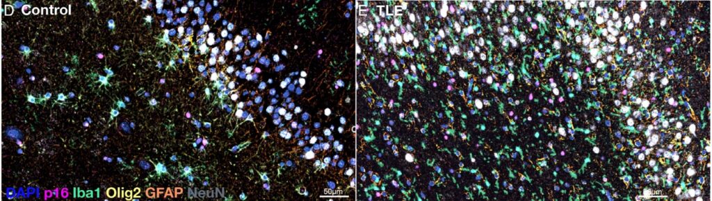
The research team examined of human hippocampal tissue obtained from individuals with medically intractable TLE. Using multiplexed immunofluorescence, the authors detected a marked increase in cells expressing canonical senescence markers compared with autopsy controls. Notably, this elevation was not restricted to a single glial population; senescence markers appeared across microglia, astrocytes, and oligodendrocytes, suggesting a widespread alteration of the glial environment within epileptic hippocampi. The magnitude of this increase exceeded what would be expected from age alone, indicating that epileptic pathology itself strongly promotes senescence. The authors turned to a pilocarpine-induced mouse model of TLE to establish temporal and mechanistic relationships. Senescence markers rose rapidly after status epilepticus, emerging within the latent period and persisting into the chronic phase. Transcript-level analyses and reporter mouse lines confirmed that this phenotype was sustained rather than transient. Although multiple cell types exhibited senescence-associated features, microglia accounted for the largest fraction of senescent cells, prompting further functional analysis of this population. The team performed imaging which showed that senescent microglia displayed consistent alterations in morphology and baseline process motility, consistent with a chronically activated yet functionally constrained state. Importantly, these changes did not simply reflect global microglial activation, as targeted depletion of non-senescent microglia failed to reproduce the observed effects on seizures or cognition. This distinction suggested that senescent microglia represent a functionally distinct subset with disproportionate pathological influence. Moreover, the authors implemented two independent senolytic strategies. In a genetic model permitting selective ablation of p16-expressing cells, partial clearance of senescent cells led to striking outcomes. Treated animals exhibited reduced seizure frequency, improved hippocampal synaptic plasticity, and normalization of spatial memory performance. Remarkably, a subset of animals was entirely protected from developing chronic epilepsy. Parallel experiments using a pharmacological senolytic cocktail achieved comparable reductions in senescent burden and similarly improved behavioral and electrophysiological outcomes.
In conclusion, the research work of Georgetown University scientists demonstrated that senescent cells accumulate in both human TLE and experimental epilepsy, and that their selective removal attenuates seizures and cognitive deficits, the study introduces senescence as a previously unrecognized driver of epileptogenesis. Epilepsy research has long focused on neurons as primary agents of pathology, with glial cells often viewed as secondary responders. The present findings challenge this hierarchy by identifying a small population of senescent glia that disproportionately influences disease trajectory. Despite representing a minority of total cells, these senescent populations appear capable of reshaping the inflammatory and synaptic landscape of the hippocampus in ways that favor seizure persistence and memory dysfunction.
Additionally, the findings suggest that disease modification in epilepsy may be achievable without directly targeting neuronal excitability. Senolytic strategies operate upstream of seizures by altering the cellular environment in which hyperexcitability emerges. This distinction is particularly important for patients with drug-resistant epilepsy, for whom conventional antiseizure medications offer diminishing returns. The partial protection from epilepsy observed in treated animals underscores the possibility that timely intervention during epileptogenesis could prevent the consolidation of chronic disease. The study also highlights the importance of cellular specificity. Broad depletion of microglia failed to improve outcomes, whereas selective elimination of senescent cells proved beneficial. This finding cautions against indiscriminate anti-inflammatory or glial-suppressive approaches and instead supports precision strategies that preserve protective functions while removing pathological subsets. The work has broader relevance to neurological disorders characterized by chronic inflammation and cognitive decline. Senescence has been implicated in neurodegenerative diseases, and the overlap in pathological mechanisms raises the possibility that senolytic therapies could exert cross-disease benefits. However, the study also emphasizes the need for careful translational consideration, as senescence plays complex roles in tissue repair and tumor suppression. In sum, the work of Professor Patrick Forcelli and colleagues opens a new therapeutic avenue with implications that extend well beyond epilepsy itself.

Reference
Tahiyana Khan, David J. McFall, Abbas I. Hussain, Logan A. Frayser, Timothy P. Casilli, Meaghan C. Steck, Irene Sanchez‐Brualla, Noah M. Kuehn, Michelle Cho, Jacqueline A. Barnes, Brent T. Harris, Stefano Vicini, Patrick A. Forcelli. Senescent Cell Clearance Ameliorates Temporal Lobe Epilepsy and Associated Spatial Memory Deficits in Mice. Annals of Neurology, 2025; DOI: 10.1002/ana.78118
Go to Journal of Annals of Neurology.