
Significance
Fluorescent probes tend to behave well under idealized conditions, but their performance often drifts once they are placed in environments that look even vaguely biological. Emission can shift and intensity can fade, while ionic strength, oxidative species, and sustained irradiation erode signal stability. Dopamine sensing only complicates this further. Dopamine is redox-active, easily oxidized, and surrounded by structurally similar small molecules in physiological fluids. It binds, rearranges, and reacts. A probe that depends on optical modulation has to tolerate all of that at once and selectivity alone is not enough; the material must remain chemically and optically intact while doing so. Achieving this balance remains challenging. Silicon quantum dots represent a promising platform for this purpose. The absence of heavy metals immediately removes a major translational barrier that persists with cadmium- or zinc-based systems. Still, the theoretical advantage of silicon has not automatically translated into usable sensors. Many published approaches lean on multi-step surface functionalization or require fairly aggressive synthetic conditions. Post-synthetic passivation is common. Each of those interventions introduces variability and reproducibility becomes an issue, especially when scaling up. Fluorescence-based dopamine detection, if it is going to be practical, should not rely on enzymatic cascades or complicated amplification schemes. Excessive engineering of the detection architecture reduces the inherent simplicity of fluorescence-based sensing, the original appeal of fluorescence as a simple readout begins to disappear. There is also the antibacterial angle, which introduces a different kind of tension. Reactive oxygen species can disrupt membranes, damage DNA, oxidize proteins. That part is well established. The difficulty is that ROS do not discriminate particularly well between bacterial and mammalian cells. Many metal and metal oxide nanoparticles generate oxidative stress effectively, but long-term biosafety is still an open question. So the problem is not whether a nanomaterial can induce oxidative damage, but whether it can do so in a way that is controlled, predictable, and compatible with surrounding tissue.
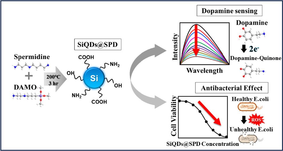
In a recent study published in ACS Applied Bio Materials, Aloke Bapli, Hyeryeong Lee, Minchae Kang, Jinmin Lee, and Professor Sang Hak Lee (Department of Chemistry, Pusan National University) addressed these challenges with a relatively restrained design. They prepared blue-emitting silicon quantum dots, denoted SiQDs@SPD, using a one-step hydrothermal reaction between spermidine and N-[3-(trimethoxysilyl)-propyl]-ethylenediamine. The appeal of this route lies in its simplicity and consolidation. Amine- and oxygen-containing surface groups form during synthesis, not afterward. That detail matters. The resulting particles disperse in water without additional modification and remain accessible for molecular interaction. The synthetic procedure itself is straightforward: spermidine dissolved in water, DAMO introduced under stirring, then heating at 200 °C for three hours in a Teflon-lined autoclave. Afterward, they removed residual precursors by centrifugation and filtration, followed by freeze-drying. Transmission electron microscopy showed particles around 2.8 nm in diameter while AFM height measurements gave an average near 3.5 nm. Additionally, the team performed surface characterization through FTIR and XPS confirmed the presence of –NH2, –OH, and other oxygenated functionalities, along with Si–C, Si–N, and Si–O bonding environments. These groups are not incidental. They define how the quantum dots interact with solvent and analytes, and they influence electronic coupling at the interface. In this system, surface chemistry and optical behavior are not separable considerations; they are structurally linked from the beginning.
The optical response is fairly well defined. In absorption, distinct features appear in the near-UV, and when the material is excited at the higher-energy band it emits in the blue region, centered close to four hundred nanometers. The quantum yield is reported to be a little under one quarter, which is respectable for a silicon system prepared this simply. Batch-to-batch reproducibility was evaluated, and overlapping spectra were observed, which at least suggests that the hydrothermal process is not drifting unpredictably from run to run. Emission does shift slightly with excitation wavelength, but not dramatically. Compared with many carbon dots, where the peak can wander significantly, these SiQDs look comparatively restrained. That would normally imply a relatively homogeneous emissive landscape, although the interaction with dopamine later complicates that interpretation.
When the authors added dopamine, the fluorescence intensity decreases progressively. Over a defined concentration window the response is linear, and the detection limit falls in the tens of micromolar range. The excitation spectrum changes and a new absorption feature grows near the emission region, consistent with formation of dopamine-quinone. Lifetimes remain almost unchanged after dopamine addition, which supports a static quenching process, likely involving ground-state complexation through surface carboxyl and hydroxyl groups. In antibacterial assays, the researchers exposed E. coli and S. aureus to increasing SiQDs@SPD concentrations. They determined minimum inhibitory concentrations of 200 μg/mL for E. coli and 25 μg/mL for S. aureus. They compared these effects with hydrogen peroxide and showed stronger bacterial suppression by the quantum dots. Through DIBF bleaching and singlet oxygen sensor fluorescence, they confirmed singlet oxygen generation. Zeta potential measurements shifted from negative values toward neutrality after SiQDs@SPD treatment, indicating surface adsorption that likely facilitates oxidative damage. The resistance-passaging experiment over six days revealed no measurable adaptation under sub-MIC exposure. That absence of resistance is notable, though long-term evolutionary pressures would require broader evaluation.
What Professor Sang Hak Lee’s and colleagues successfully demonstrated the new hydrothermal route is convenient synthesis and folds surface functionalization into the growth stage itself. That matters. Amine and oxygen-containing groups become part of the particle as it forms, rather than something appended later. As a result, the dots disperse in water and still retain chemically accessible sites. When dopamine oxidizes to its quinone form, its electron-accepting character can couple with those surface states. The fluorescence decreases without a meaningful shift in lifetime, which is consistent with static quenching. In other words, the sensing response seems to arise from the inherent surface chemistry, not from engineered reporters layered on top. The antibacterial data add another layer. Under light, the particles generate singlet oxygen, and zeta potential changes suggest that they adsorb onto bacterial surfaces. That combination of physical proximity plus oxidative stress can drive the suppression of growth. The lower inhibitory concentration observed for Gram-positive strains probably reflects structural differences in the cell envelope. Repeated exposure below the inhibitory threshold did not yield detectable resistance during the study window. That observation is encouraging, although it would be premature to assume long-term stability under clinical pressures. From a medical standpoint, the innovation is in that neurotransmitter sensing and antimicrobial activity coexist in a single silicon-based material. Dopamine imbalance is implicated in several neurological conditions, and probes that remain optically stable in complex media reduce practical obstacles to detection. At the same time, antibiotic resistance continues to narrow therapeutic options. A system that couples selective dopamine responsiveness with light-activated antibacterial behavior suggests, a combined diagnostic and therapeutic platform.


Reference
Bapli A, Lee H, Kang M, Lee J, Lee SH. One-Step Hydrothermal Synthesis of Silicon Quantum Dots for Dopamine Detection and Their Antibacterial Activity against Escherichia coli and Staphylococcus aureus. ACS Appl Bio Mater. 2025;8(8):7023-7036. doi: 10.1021/acsabm.5c00752.
Go to ACS Applied Bio Materials.