Significance
A central element in this story is ELOVL2, an elongase whose promoter is one of the most reliable molecular clocks of aging in mammals. Its activity converts 22:5n-3 into 24:5n-3, a precursor from which the retina derives a suite of VLC-PUFAs that ultimately shape the membrane environment of photoreceptor outer segments. Prior work hinted that ELOVL2 expression declines with age and that its methylation state tracks chronological aging in retina, liver, and blood. Yet the functional implications of progressive ELOVL2 downregulation for retinal performance had remained uncertain. Observational studies also pointed toward a relationship between lower retinal DHA abundance and increased AMD risk, but oral supplementation trials produced inconsistent outcomes, perhaps because nutritional routes fail to deliver adequate amounts of VLC-PUFAs directly to the eye.
To this account, new research paper published in Science Translational Medicine and led by Professors Vladimir Kefalov, Dorota Skowronska-Krawczyk Fangyuan from the University of California, Irvine and contributed by Gao, Emily Tom, Cezary Rydz, William Cho, Alexander Kolesnikov, Yutong Sha, Anastasios Papadam, Samantha Jafari, Andrew Joseph, Ava Ahanchi, Nika Balalaei Someh Saraei, David Lyon, Andrzej Foik, Qing Nie, and Felix Grassmann, researchers developed two complementary models: an aging-retina model that maps the natural decline of VLC-PUFAs and a genetic ELOVL2-deficiency model that accelerates these biochemical and functional losses.
The investigators first mapped age-dependent lipid changes by performing LC–MS lipidomics on mouse retinas spanning 3 to 23 months. The data revealed pronounced reductions in DHA and multiple VLC-PUFA species in older retinas, accompanied by shifts in phosphatidylcholine and phosphatidylethanolamine abundance and altered membrane-associated lipid ontology signatures. When photoreceptor outer segments were purified, the decline in VLC-PUFA–containing phospholipids became even more striking, implying that membrane composition itself drifts toward a less supportive milieu for phototransduction Behavioral and electrophysiological assays reinforced this biochemical portrait: older mice showed reduced contrast sensitivity, diminished scotopic and photopic ERG amplitudes, slower dark-adaptation kinetics, and attenuated oscillatory potentials. Immunostaining demonstrated increased deposits of APOE and C3d beneath the RPE, underscoring that molecular hallmarks of aging accompany functional decline.
To test whether ELOVL2 loss accelerates these features, the group examined Elovl2C234W mice lacking enzymatic activity. Lipidomics revealed a selective depletion of products derived from ELOVL2, closely mirroring the aging lipid profile. These mutants displayed substantial deficits in rod function by 12 months, resembling wild-type animals six months older. Yet retinal thickness remained intact, demonstrating that functional deterioration precedes discernible degeneration. Transcriptomic comparisons showed that Elovl2-deficient retinas adopt gene-expression patterns characteristic of aging, including activation of synaptic remodeling, TORC1 signaling, and stress-response pathways.
The researchers then tested whether retinal function could be restored by directly supplying the missing lipid. They injected 24:5n-3, the immediate product of ELOVL2, into the vitreous of aged mice. Dose-response testing identified 0.36 nmol as optimal. Five days after injection, ERGs showed robust improvements in scotopic and photopic amplitudes, and visually evoked potentials recorded from the superior colliculus increased markedly. Comparable supplementation with DHA, EPA, or other related fatty acids failed to elicit similar recovery, indicating a remarkable specificity for 24:5n-3. Lipid analysis confirmed heightened incorporation of VLC-PUFA–containing phospholipids into photoreceptor membranes after treatment. RNA sequencing revealed downregulation of complement, oxidative stress, microglial activation, and inflammatory networks, accompanied by reduced C3d and APOE deposition in the RPE. Long-term testing demonstrated that functional improvement persisted for several weeks after a single dose, though repeated injections gradually produced diminishing returns. Finally, genetic analyses in human cohorts established that variants within ELOVL2 correlate with earlier AMD onset, strengthening the translational relevance of the findings.
In conclusion, the new study by Professor Dorota Skowronska-Krawczyk Fangyuan and colleagues developed new models that demonstrated that insufficient production of 24:5n-3 is a central driver of retinal aging. By supplementing this lipid intravitreally, the researchers established a targeted molecular therapy capable of reversing several hallmarks of age-related visual decline. Indeed, the new work reshapes how retinal aging is conceptualized, illustrating that the slow erosion of VLC-PUFA abundance is not a passive biomarker of senescence but a modifiable determinant of visual capability. By connecting biochemical erosion, physiological decline, and molecular reprogramming, the authors provide a mechanistic account that links membrane lipid composition with the integrity of photoreceptor signaling. The discovery that ELOVL2 activity not only predicts chronological age but actively regulates the functional resilience of the retina highlights a metabolic axis that had been overlooked in therapeutic design. Their evidence shows that photoreceptors do not merely suffer downstream consequences of aging; they experience a progressive insufficiency of specific lipids essential for sustaining rapid signal transduction.
The therapeutic implications are substantial. Dietary n-3 PUFA interventions, though appealing, cannot deliver sufficient quantities of VLC-PUFAs to the eye. The present work shows that intravitreal replacement bypasses this limitation. The ability of a single molecule—24:5n-3—to restore contrast sensitivity, enhance photopic and scotopic responses, shorten dark-adaptation delays, and reduce sub-RPE inflammatory deposits demonstrates that retinal aging retains an unexpected degree of reversibility. Molecular analysis revealed a partial reversion of inflammatory and complement-related transcriptional signatures, suggesting that membrane lipid restoration influences circuit-level and innate immune pathways. This interplay between lipid metabolism and microglial tone echoes the growing view that age-related vision loss is shaped jointly by metabolic strain and chronic inflammation. Equally important is the connection to human disease. The association between ELOVL2 genetic variants and earlier onset of intermediate AMD strengthens the biological narrative and positions ELOVL2 not simply as a metabolic enzyme but as a potential therapeutic entry point. Although the translation of intravitreal PUFA therapy to humans requires careful evaluation, the demonstration that a single elongation product can modulate both functional and molecular hallmarks of aging challenges long-standing assumptions that early AMD is refractory to metabolic intervention. Moreover, the study suggests that metabolic rejuvenation targeting the precise molecules whose decline destabilizes cellular systems could complement existing strategies for neuroprotection and tissue maintenance. The retina, with its remarkable sensitivity to lipid composition, proves to be an ideal model for testing such metabolic interventions. What emerges is a therapeutic rationale grounded in molecular precision, pointing toward interventions that could meaningfully delay or prevent age-related vision loss.
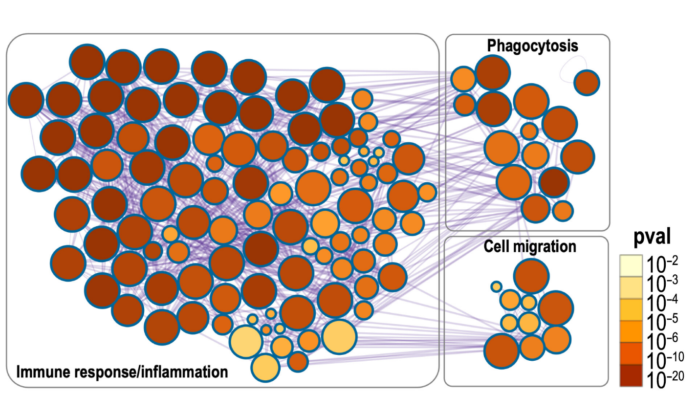
Figure: Metascape analysis demonstrated down-regulation of immune response, inflammation, microglial phagocytosis, and cell migration pathways after 24:5n-3 supplementation. The network plot visualizes functionally grouped enriched terms, where each node represents a biological pathway or process. Node size reflects the number of genes associated with each term, and node color indicates statistical significance, with darker colors representing lower P values. Image credit: Science Translational Medicine, 2025; 17 (817) DOI: 10.1126/scitranslmed.ads5769